dsGreen is a less toxic alternative to Ethidium bromide. The dsGreen dye is a highly sensitive stain used for visualizing dsDNA in agarose and polyacrylamide gels.
dsGreen has several advantages compared to the routinely used Ethidium bromide:
✔ 25-fold more sensitive for dsDNA and 50-fold for RNA detection
✔ Much less mutagenic
✔ Can be used with either blue light or UV excitation
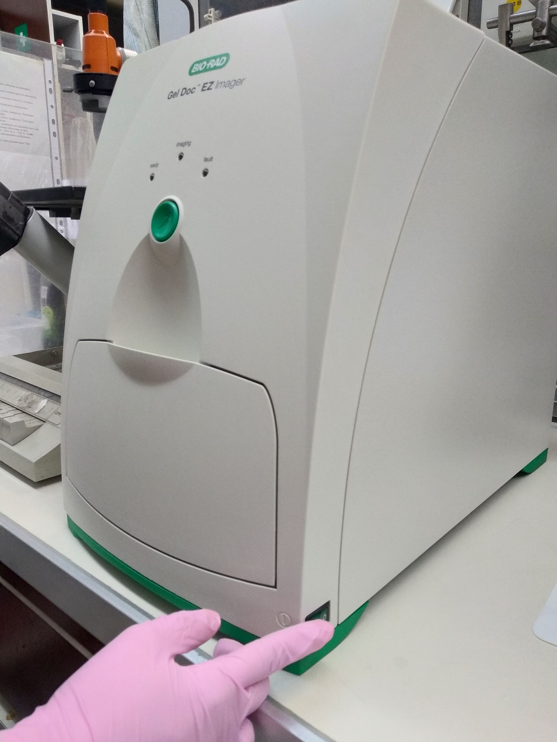
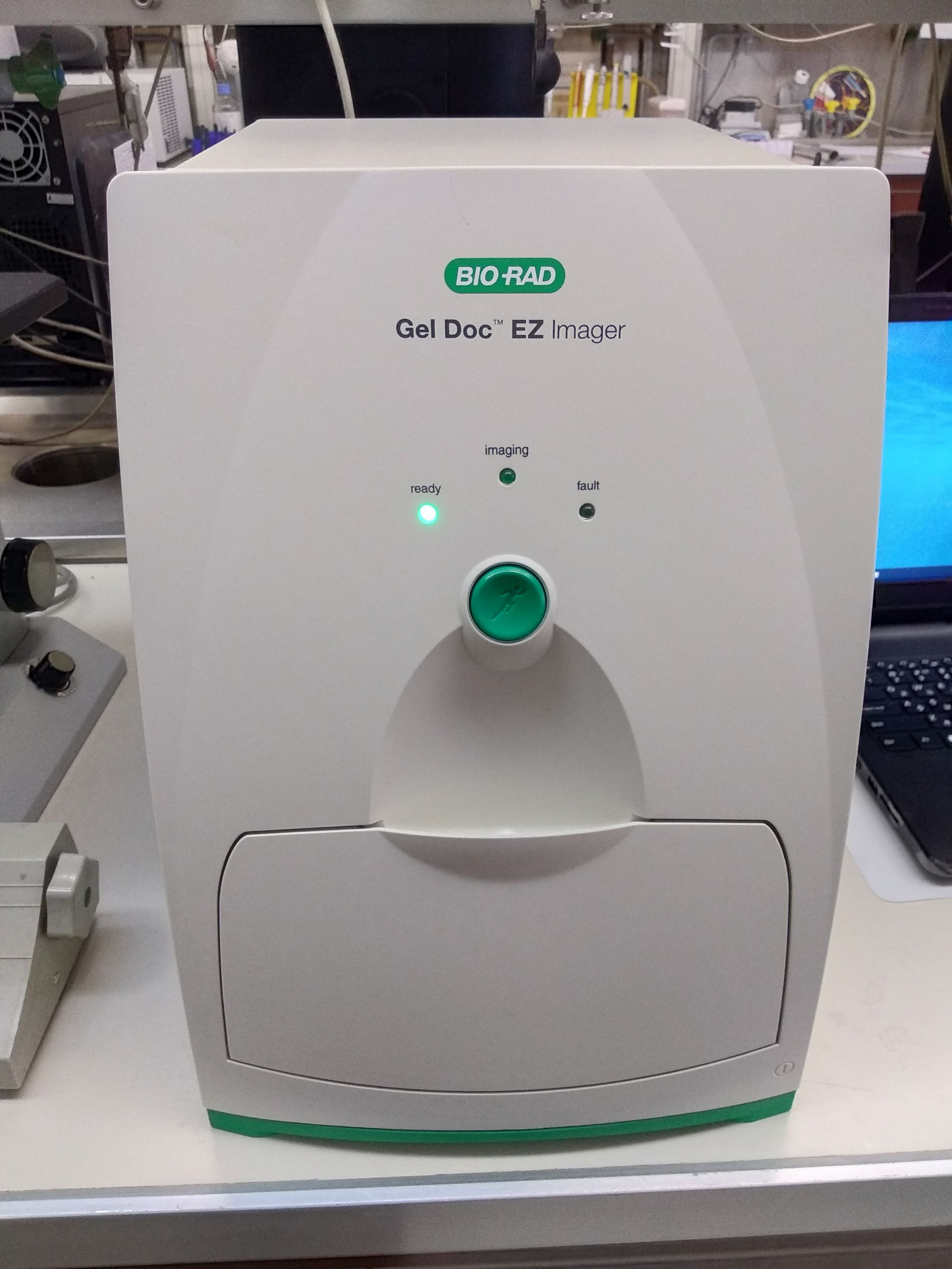
Among others, Gel Imaging Systems supplied by Bio-Rad are suitable for visualizing gels stained with dsGreen. The image of the gel stained with dsGreen should be taken using
· the XcitaBlue Conversion Screen for Gel Doc™ XR+ System
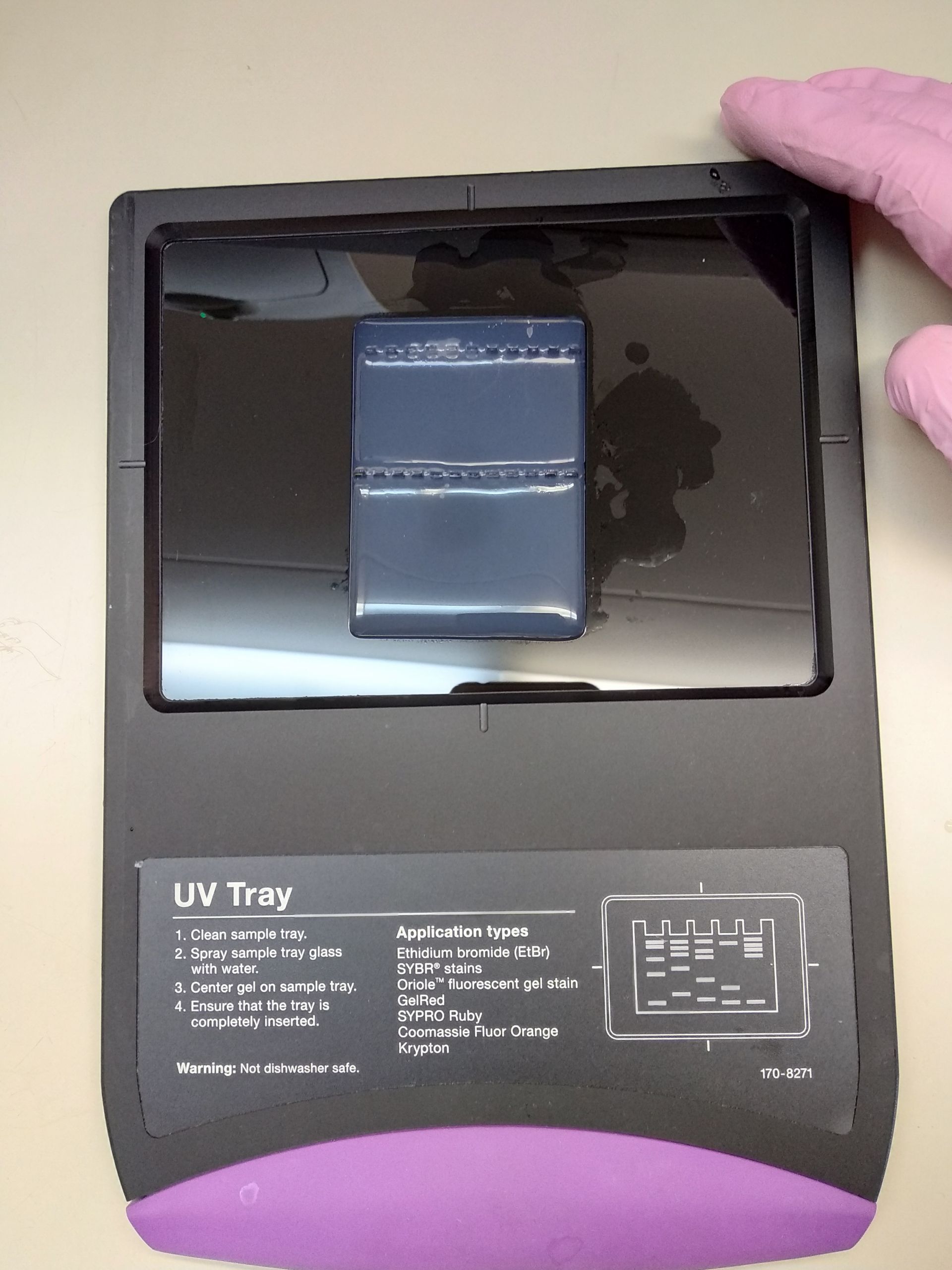
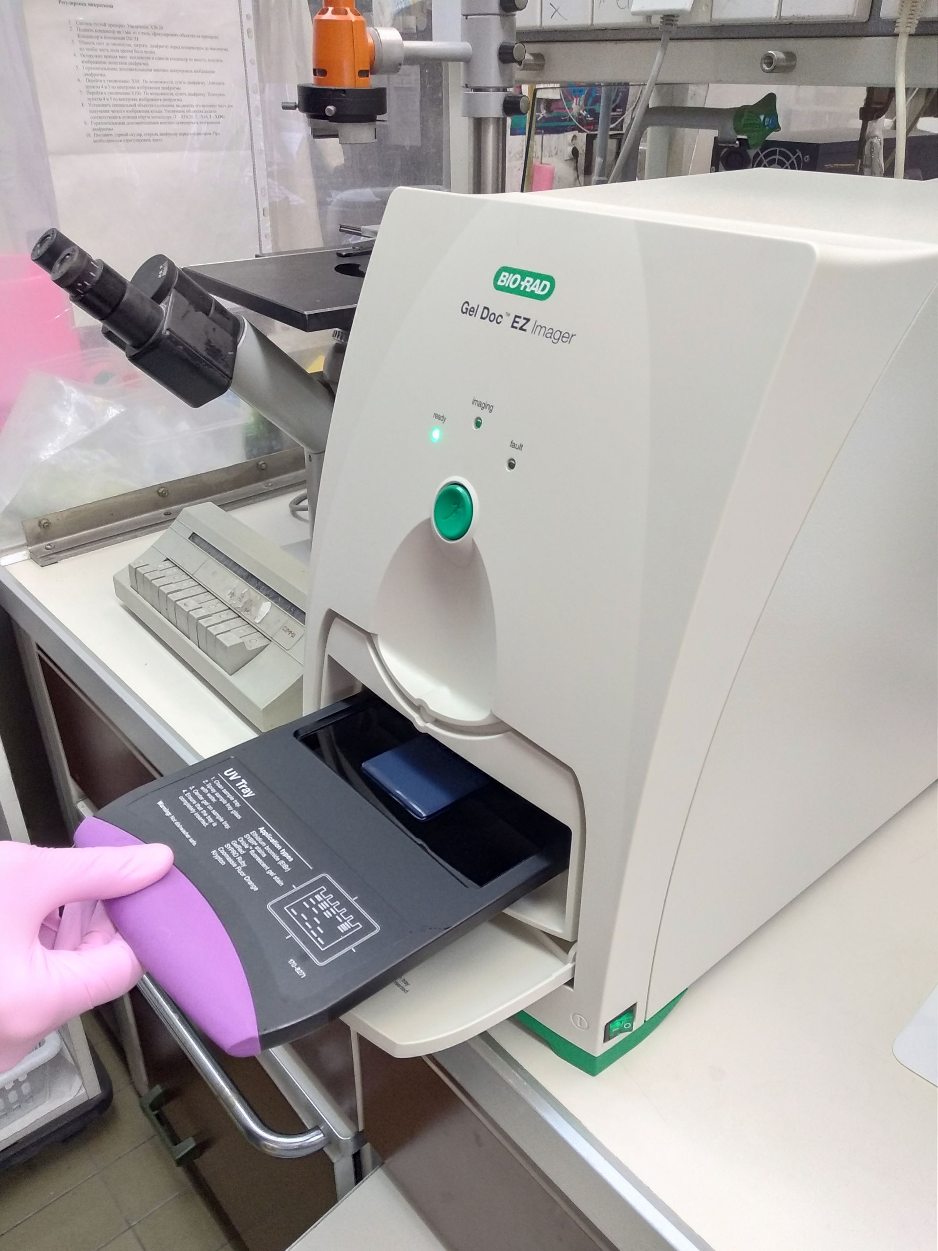
· UV Tray or Blue Tray for Gel Doc™ EZ System.
As a first step gel should be stained with dsGreen. There are three methods to stain a gel with dsGreen. Still, we recommend the gel soaking method, which has been shown to be the most effective in terms of DNA fragment separation quality and analysis sensitivity. After the gel has been stained with dsGreen, it will take you a few minutes to visualize the gel.
The Gel Doc EZ™ System offers numerous options for advanced users, including creating default protocols, analysis tools, and quantity tools. In this article, we demonstrate a simple method for capturing an image of the gel stained with dsGreen. For detailed information about Bio-Rad's Gel Imaging Systems, refer to the manufacturer's documentation.
Step 3.
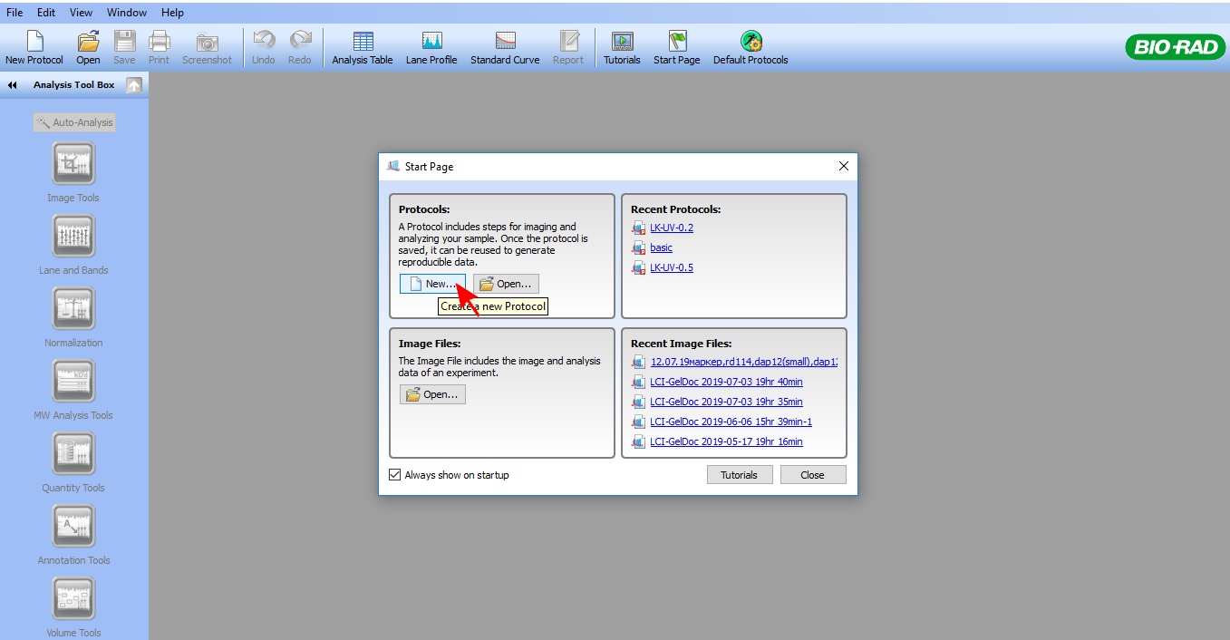
On the toolbar, click New.
*If you have created a default protocol, press the green Run button on the front of the imager. The default protocol runs automatically.
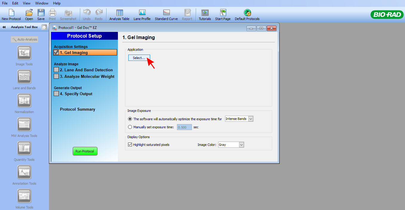
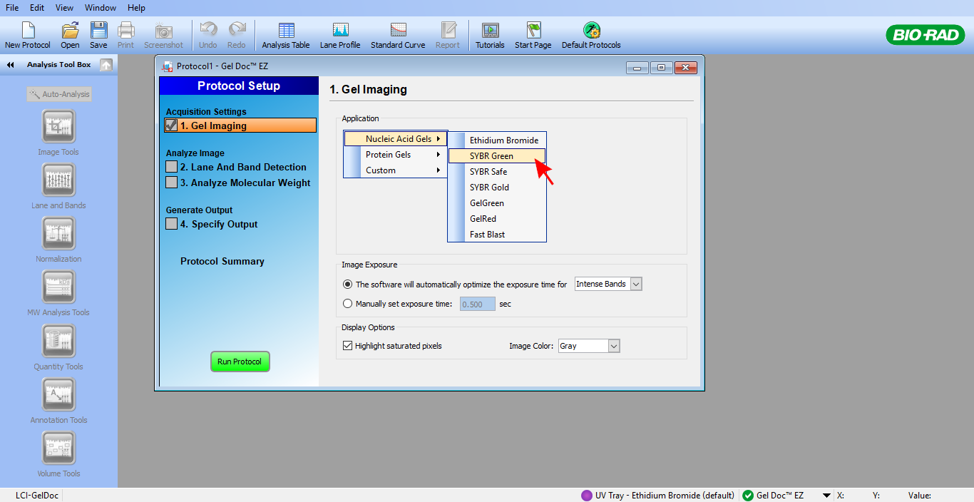
In the Gel Imaging pane, click Select and choose Nucleic Acid Gels -> SYBR™ Green.
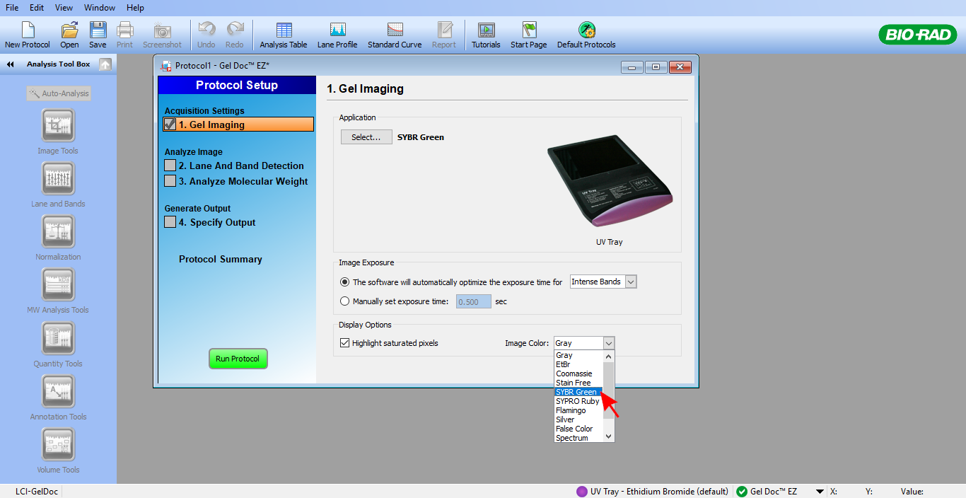
As an Image Color, select SYBR™ Green.
You can view the image with any color scheme, but the SYBR™ Green color scheme makes it easier to view naturally.
Click Run Protocol. You will see the imaging run progress bar.

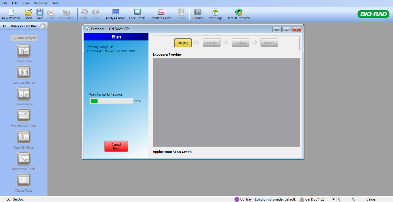
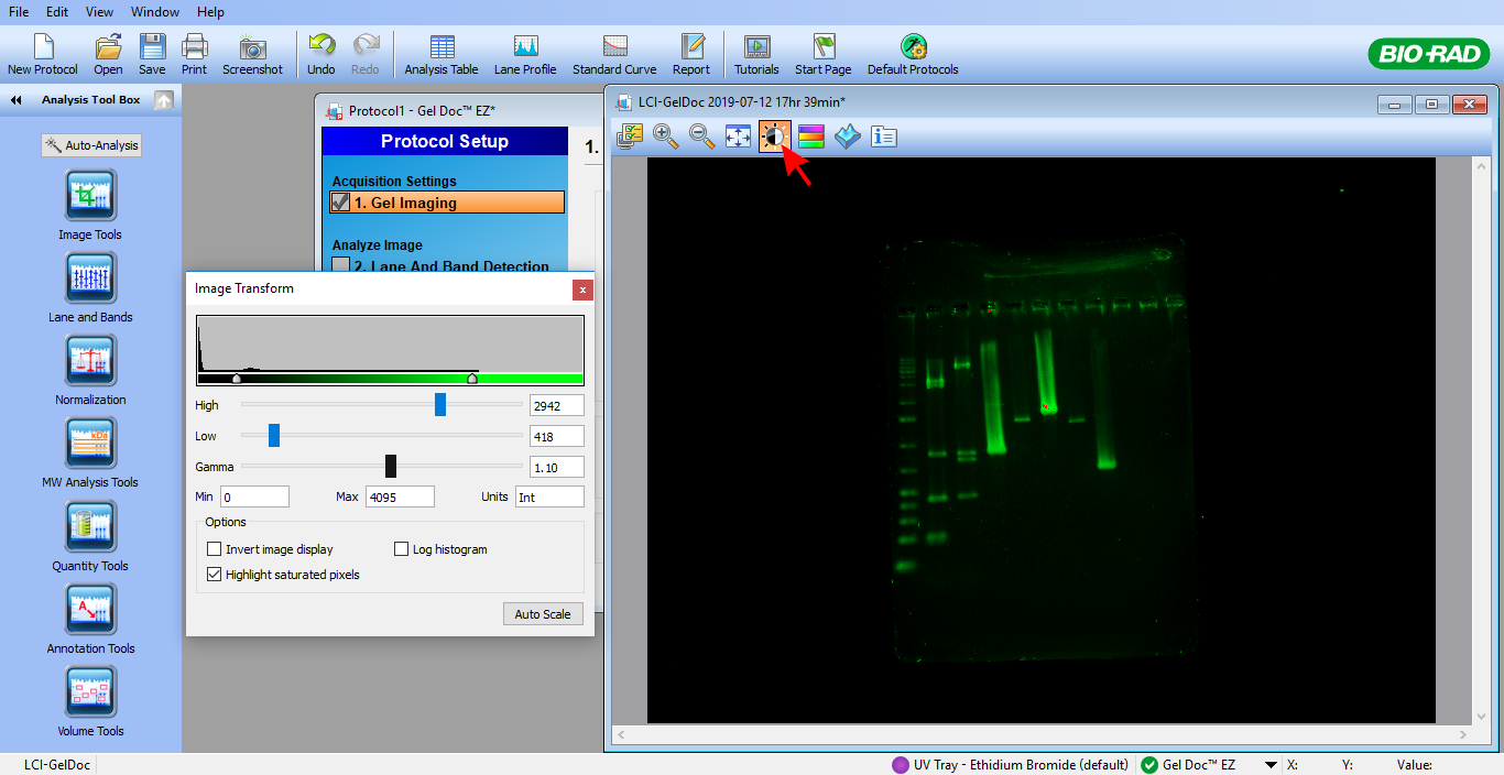
A new window will appear, and you will see the image of your sample gel.
In this window, you can process the image.
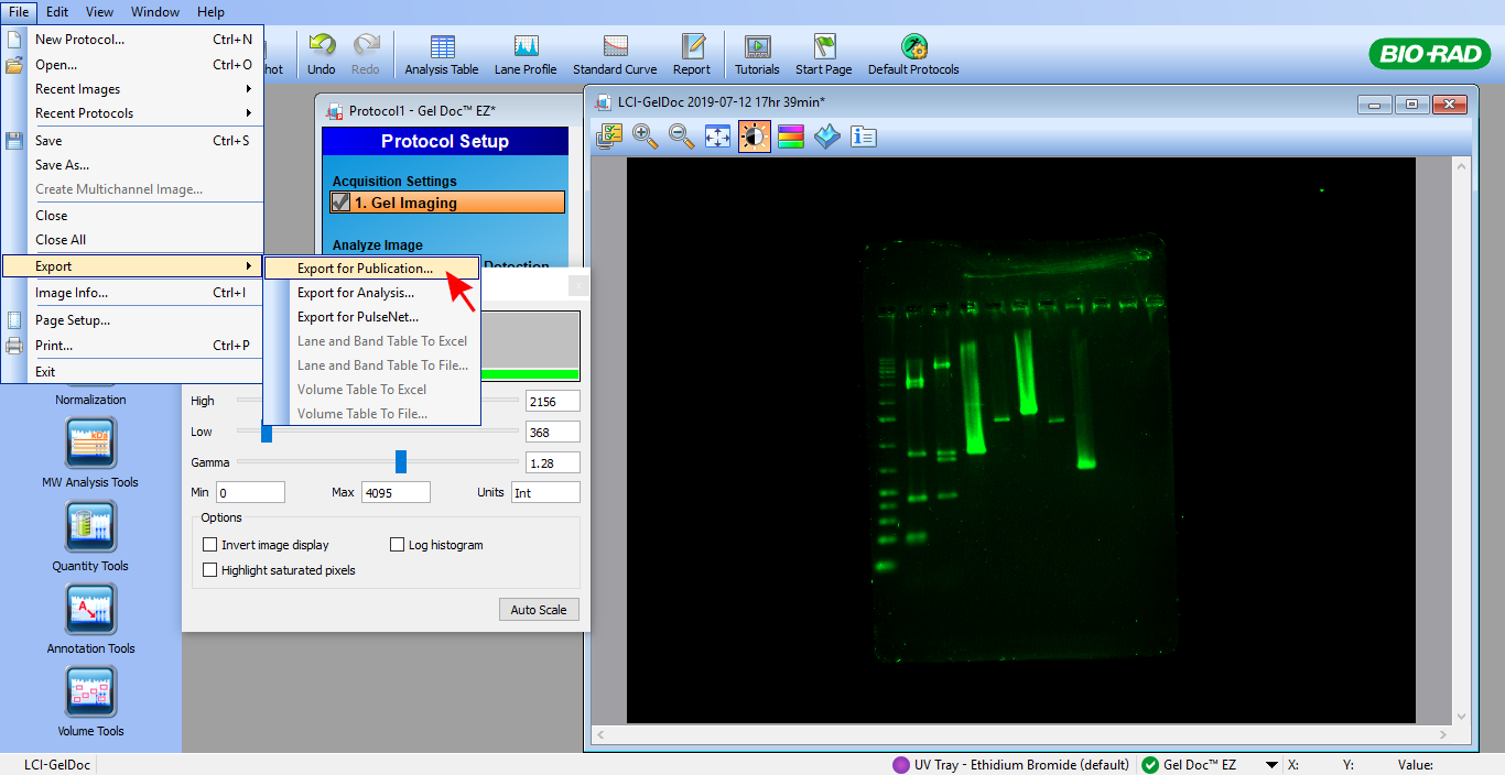
To export the image, click File -> Export -> Export for Publication.
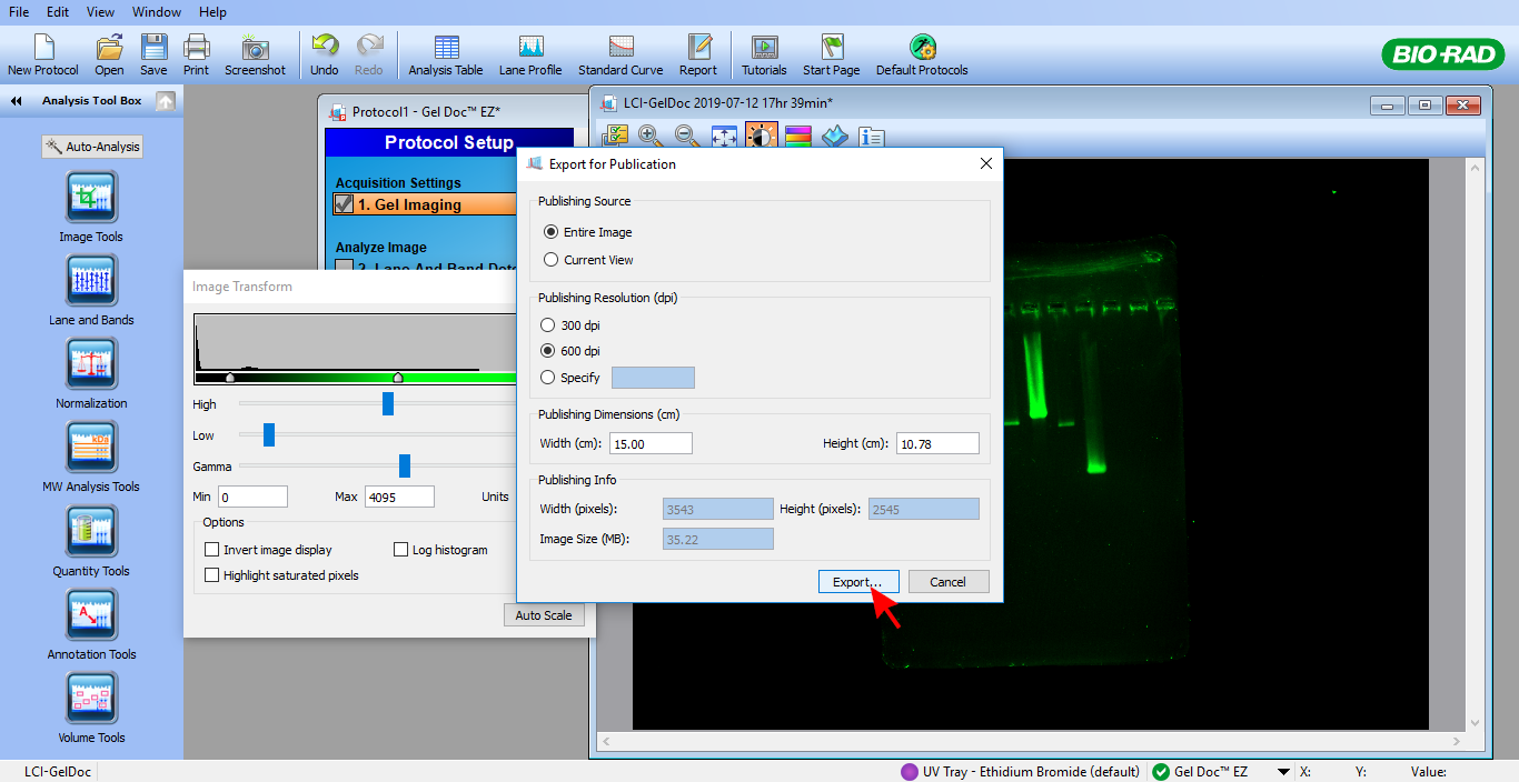
Select the resolution and dimensions. Click Export and save the image in the folder of your choice.
SYBR™ is a trademark of Thermo Fisher Scientific.
Gel Doc™ is a trademark of Bio-Rad Laboratories.

