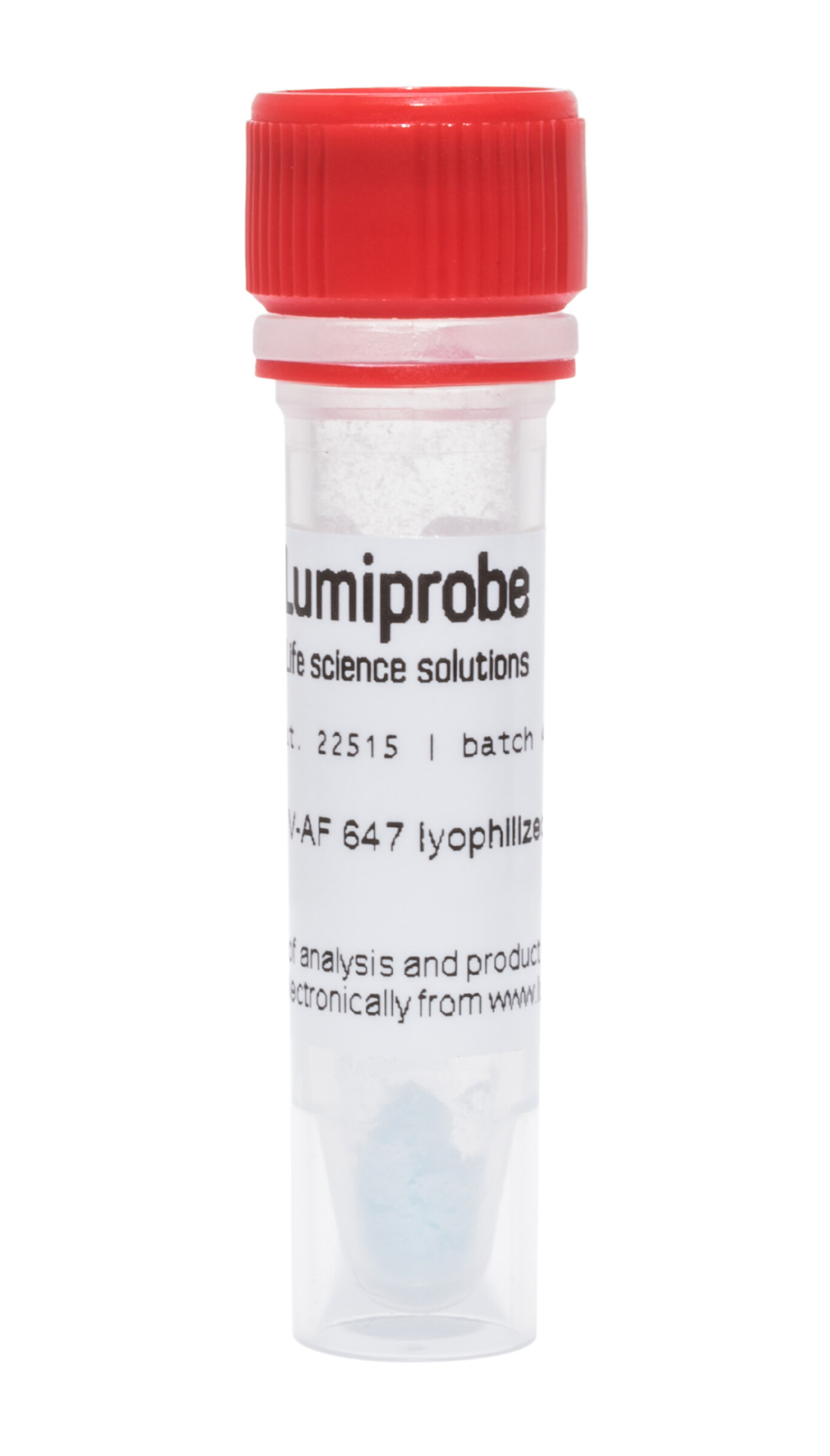
Annexin V, AF 647 conjugate
| Cat. # | Quantity | Price | Lead time | Buy this product |
|---|---|---|---|---|
| 12515 | 1 ug |
–
|
in stock | |
| 22515 | 5 ug |
$119.00
|
in stock |


Annexin V (or Annexin A5) belongs to the phospholipid-binding Annexin family of intracellular proteins. In flow cytometry and fluorescent microscopy, Annexin V is commonly used to detect apoptotic cells by its ability to specifically bind to phosphatidylserine, which migrates from the inner to the outer leaflet of the plasma membrane during the early stages of apoptosis.
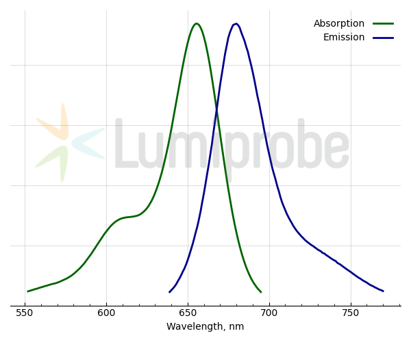
This Annexin V is a lyophilized conjugate with AF 647, a bright, photostable, hydrophilic far-red fluorophore with spectral characteristics similar to Cyanine5 (absorption max. at 655 nm, emission max. at 680 nm). Far-red fluorescent tags with excitation above 600 nm and emission further than 650 nm are valuable for imaging techniques because of the lower background autofluorescence at these wavelengths.
Annexin V AF 647 staining alone does not segregate the populations of apoptotic and necrotic cells. For this, additional staining with nuclear dyes that do not penetrate living cells — with propidium iodide or YODi-3 is required. For this purpose, you can also use our ready-to-use Annexin V AF 647 apoptosis detection kit.
Dissolve the content of the Annexin V AF lyophilized conjugate tube in 50 µL (12515) or in 250 µL (22515) of deionized water.
Important! The dissolved conjugate should be stored protected from light at


Absorption and emission spectra of AF 647
Customers also purchased with this product


")


General properties
| Appearance: | blue solid |
| Solubility: | good in water |
| Storage conditions: | Store at -20 °С for 9 months from date of receipt. Transportation: at room temperature for 1 week. |
| MSDS: | Download |
| Product specifications |
Spectral properties
| Excitation/absorption maximum, nm: | 655 |
| Emission maximum, nm: | 680 |


 $
$ 
