Deep-Red Fluorescent Nissl Stain
| Cat. # | Quantity | Price | Lead time | Buy this product |
|---|---|---|---|---|
| 1A010 | 50 uL |
–
|
in stock | |
| 2A010 | 500 uL |
$139.00
|
in stock |


Nissl staining is a commonly used histological technique to visualize neural tissue morphology. The method is based on the interaction of basic dyes with the nucleic acid content of cells. Due to intensive protein synthesis, the perikarya of neurons has abundant ribosomal RNA in the rough endoplasmic reticulum (’Nissl substance’), and cytoplasmic staining of neurons is much stronger than in nuclei. On this basis, stained neurons can be distinguished from glial cells, and therefore, Nissl staining is considered specific to detect neurons.
We offer highly concentrated (1,000×) Fluorescent Nissl Stains with different spectral properties.
Deep-Red Fluorescent Nissl Stain is a cell-impermeant dye that is nonfluorescent in the absence of nucleic acids but exhibits a significant fluorescence enhancement upon binding to RNA and DNA. The long-wavelength fluorescence of Deep-Red Fluorescent Nissl Stain is well separated from green and red fluorophores, which makes it ideal for multicolor fluorescence labeling experiments.

Product in action
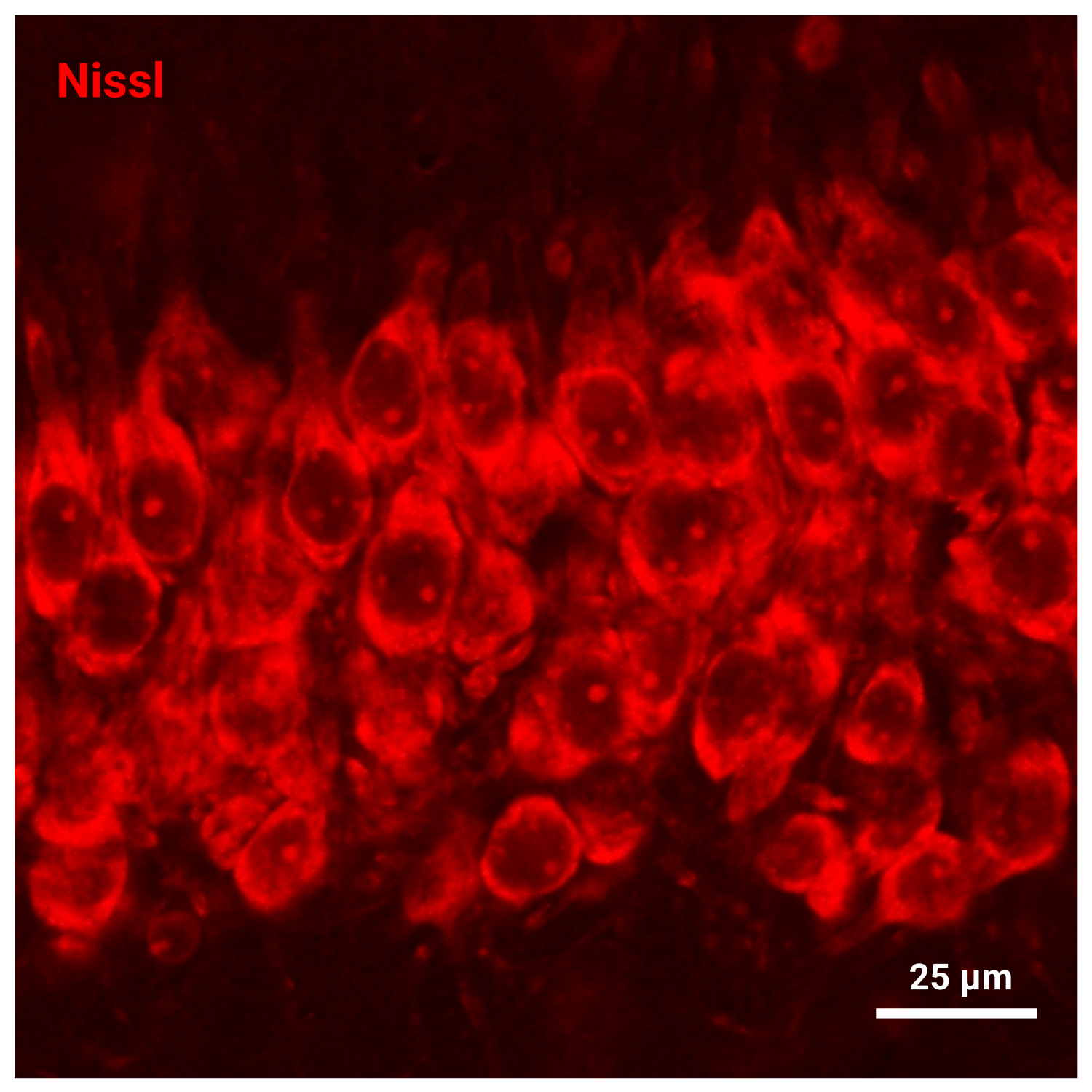
Neurons of the CA3 area of the hippocampus imaged using Deep-Red Fluorescent Nissl Stain

Mouse brain section stained with Deep-Red Fluorescent Nissl Stain
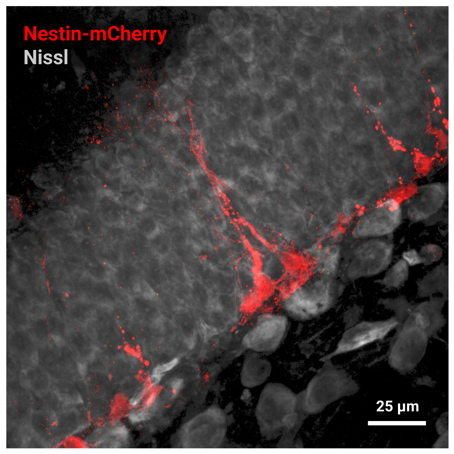
Dentate gyrus of Nestin-mCherry mouse stained with Deep-Red Fluorescent Nissl Stain
Excitation and emission spectra of Deep-Red Fluorescent Nissl Stain

Recommended protocol
Calculator
Customers also purchased with this product

")


General properties
| Appearance: | blue liquid |
| Quality control: | NMR 1H and HPLC-MS (95+%) |
| Storage conditions: | 24 months after receival at -20°C in the dark. Transportation: at room temperature for up to 3 weeks. Desiccate. |
| MSDS: | Download |
| Product specifications |
Spectral properties
| Excitation/absorption maximum, nm: | 644 |
| Emission maximum, nm: | 662 |


 $
$ 
